

Radiology
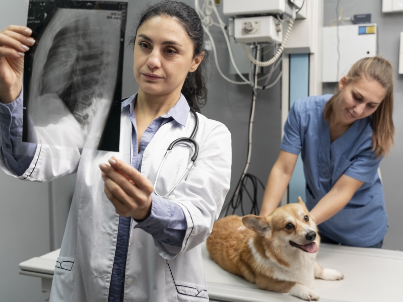
All X-rays taken at Devashrya Animal Foundation are done on-site. We employ digital radiology because it consumes less radiation and enables us to respond to you more quickly. These examinations can reveal oral problems, some soft tissue damage, skeletal fractures, and foreign substances.
Even while X-rays are non-invasive, they do demand that the patient stay still, so your pet may need to be sedated or given anesthetic. However, sedation is typically only temporary, and patients normally recover soon.
Common Conditions & Procedures
- Abdominal, musculoskeletal, and thoracic radiography
- Myelography, excretory urography and cystography, esophageal and tracheal fluoroscopy, barium gastrointestinal investigations, and arthrography are examples of special radiographic techniques.
- Abdominal, thoracic, and musculoskeletal ultrasound examinations, ultrasonography guidance for biopsies and needle aspirations.
- Thyroid investigations & bone scans using nuclear medicine
- Assessment of portosystemic shunts, hyperthyroidism, and renal problems
- CT scans: evaluation of conditions affecting the bones, joints, heart, lungs, abdomen, head, spine, and ears. CT can be used to direct needle biopsies as necessary.
- Small animal neurologic imaging (brain and spine) and equine musculoskeletal and head imaging are both examined using MR technology.
Best Of Facilities
Our clients and their companion animals receive thorough referral services in a polite and effective manner from our fully staffed imaging and treatment center and MRI facility. Digital radiography, fluoroscopy, ultrasonography, multislice helical computed tomography, and 1.5T magnetic resonance imaging are some of the diagnostic imaging methods we can use (MRI).